Brain scans are done to help investigate the cause of persistent headache, confusion, fits, memory problems, or focal weakness in the arms or legs. They can help in confirming or ruling out the presence of lesions (e.g. toxoplasmosis, non-Hodgkin's lymphoma) or central nervous system disease (e.g. progressive multifocal leukoencephalopathy, or PML).
Brain scans are performed using magnetic resonance imaging (MRI), computed tomography (CT) or computed axial tomography (CAT), positron emission tomography (PET), and single photon emission computed tomography (SPECT). These techniques directly or indirectly image the structure and function of the brain. Structural imaging is used to diagnose tumours or injury. Functional imaging is used to diagnose metabolic diseases and lesions (such as Alzheimer’s).
In HIV care, MRI and CT/CAT scans are mainly used and the choice is often dictated by available hospital facilities and cost. Both methods are non-invasive and painless, as are PET and SPECT.
MRI uses magnetic fields and radio waves to produce brain structure images without the use of radiation. It renders brain surface and subsurface images with a high degree of anatomical detail. Originally, MRIs could only provide information on the physical appearance of the brain, including assessment of water content, inflammation, and bleeding. Functional MRI (fMRI) imaging provides the same structural information, but functioning (brain activity) information as well. MRIs can produce cross-sectional images in any direction. Some people feel claustrophobic in an MRI machine. It helps if the patient closes their eyes while the 'bed’ is sliding into the MRI tube. Some people are more comfortable if they are first given some type of sedation.
CT or CAT scans take a series of X-rays from many angles. Normally, this technique is used for viewing brain injuries, assessing ventricle size, and evaluating tissue damage caused by swelling. The scan takes just a few minutes to complete.
PET measures emissions from radioactively labelled metabolically active chemicals that have been injected into the bloodstream. When these chemicals reach the brain, two- and three-dimensional images are produced that map neurotransmitter activity. This test can also be completed in moments and is useful in the diagnosis of tumours, strokes, and other diseases that can cause dementia.
SPECT is somewhat similar to PET, but uses gamma ray emitting radioisotopes and a camera to record data. An injection of radioactive tracer is taken up by the brain and cerebral blood flow is reflected. SPECT is mainly used for imaging epilepsy or diagnosing brain disease, such as dementia.

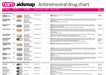 Antiretroviral drug chartA one-page reference guide to the anti-HIV drugs licensed for use in the UK or European Union, with information on formulation, dosing, key side effects and food requirements.
 Your next stepsA booklet with information for people who’ve just found
out they have HIV.
Your next stepsA booklet with information for people who’ve just found
out they have HIV.
 Coming soon: news from IAS 2019 05 July 2019The 10th International AIDS Society
conference (IAS 2019) is being held...
Coming soon: news from IAS 2019 05 July 2019The 10th International AIDS Society
conference (IAS 2019) is being held... Those who are not seen are not heard 01 July 2019A commentary on Optimising the Impact of Key Population Programming...
 The fight for access to abortion care in Europe 27 June 2019Access to
sexual and reproductive health and rights includes safe and...
The fight for access to abortion care in Europe 27 June 2019Access to
sexual and reproductive health and rights includes safe and...