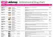
Exposure and primary infection

HIV is not able to penetrate intact skin, but can enter the bloodstream through a puncture wound, cut, or sore. HIV can also penetrate mucous membranes in the rectum, the female genital tract, the underside of the foreskin in uncircumcised men, the anus, and the urethra (the tube that carries urine). HIV is present in blood, semen, vaginal fluids, rectal mucous and breast milk.

It is not clear whether HIV is infectious as free virus outside cells or as HIV within CD4 T-cells passing to another person. The first cells to become infected with HIV are usually dendritic cells in the linings of the rectum and sexual organs. There is good evidence that other sexually transmitted diseases can increase the risk of HIV transmission, by increasing the number of immune cells in genital secretions or by causing ulceration.

In sexual transmission, HIV is trapped in the tissue of the rectum or sexual organs by dendritic cells and carried to the lymph nodes where it is presented to and recognised by a few T- and B-cells. Under T-cell control, the B-cells produce clones of plasma cells, which can release antibodies that specifically attack HIV in the bloodstream. The appearance of these antibodies against HIV antigens in the blood is called seroconversion. HIV viruses outside cells can become coated with antibodies and may then be attacked and destroyed by phagocytic cells. However, HIV makes small errors when it reproduces itself, resulting in a large amount of variability in the 'envelope antigens' on its coat. This variability allows HIV variants to escape recognition by specific antibodies and to persist in the body.

Early detection of HIV

Several different tests can be used to establish whether a person is in the early stages of HIV infection . PCR viral load is the most sensitive test for detecting HIV infection in seroconversion, though greater sensitivity may be achieved when used in conjunction with the proviral DNA test.1,2

The quantitative polymerase chain reaction (PCR) test is performed by doing a routine blood draw. PCR amplifies genetic material (RNA) and looks for actual virus by using the reverse transcriptase (RT) enzyme to multiply HIV gene sequences in the blood sample so that they show up more easily. A chemical reaction marks the virus and these markers are then measured and used to calculate the amount of virus in the bloodstream. This test is very reliable for detecting HIV in someone recently exposed to virus and will be highly accurate within 48 to 72 hours. An ultra-sensitive version of the RT-PCR test can detect as few as 50 copies/ml.

A qualitative PCR test, known as the PCR-DNA test, looks for the presence of virus, but does not measure the amount found. This is a useful test for detecting infection in infants because it will detect virus before viral load is present, but it is a more expensive test.

The branched DNA (bDNA) assay also detects the amount of virus and has results comparable to RT-PCR. The bDNA test contains a material that gives off light when it connects with HIV particles. The amount of light is measured and converted to a viral count.

In testing, high sensitivity means that a test will detect all cases of disease, but perhaps also cases where disease is not actually present. This type of error is called a 'false positive'. High specificity means that the criteria for detecting disease are very specific, and occasionally, some cases of disease will be missed. This type of error is called a 'false negative'. Most tests are a balance between these two qualities; the balance being determined by whether the priority is finding all disease or definitively ruling out disease.

Antibody testing, which is commonly used to diagnose HIV infection, is unlikely to detect HIV antibodies until at least six weeks after infection in most cases. The HIV antibody test is known as an enzyme-linked immunosorbent assay (ELISA) test.

See Types of viral load tests in Monitoring the immune system for further information on specific viral load assays.

Seroconversion illness

Seroconversion typically occurs two to twelve weeks after infection and may be associated with a flu-like illness. The most commonly reported symptoms include fever, rash, aches and pains, oral ulcers, sore throat, fatigue, and nausea.

Between 50 and 80% of people experience some or all of these symptoms at seroconversion. A smaller proportion may develop diarrhoea, ulceration of throat or genitals, anorexia, or swollen lymph nodes. Weight loss, abdominal pain, loss of appetite, oral thrush, and neurological abnormalities have also been reported as symptoms of seroconversion.

A study of seroconverters in Zambia suggested a slightly different profile for seroconversion illness. There, malaria, diarrhoea, swollen glands, inflammation, night sweats, and weakness were associated with HIV seroconversion.3 

Increased duration and severity of illness was associated with a higher viral load during seroconversion in one Kenyan study of 74 female prostitutes. Those who were asymptomatic at seroconversion had a median viral load of 216 copies/ml, those with one symptom had a median viral load of 32,111 copies/ml, and those with five or more symptoms had a median viral load of approximately 4,000,000 copies/ml.4

A US study of 170 patients found that a high initial viral load was associated with more symptoms during primary infection; however, only the initial viral load (and not the number of symptoms) was strongly correlated to the viral load set point.5 Investigators thought that aside from initial viral load, innate immune factors and/or HIV viral factors may also have contributed to determining the viral load set point.

Immune response during primary infection

The presence of high levels of HIV in the body stimulates the immune system and huge numbers of HIV-specific CD4 T-cells are produced in an attempt to contain the virus. However, these activated CD4 T-cells are also prime targets for the virus and can be rapidly infected and destroyed, leaving the body with weakened anti-HIV immune responses.

Long-term non-progressors tend to maintain high levels of HIV-specific CD4 T-cells and CD8 T-cells that effectively control HIV replication and keep viral loads low. Stronger HIV-specific CD8 T-cell responses are significantly associated with greater viral load decline during primary infection.6 CD8 T-cells act against HIV in primary infection by killing HIV-infected cells and secreting chemokines.

HIV-specific CD8 T-cells recognise a specific genetic sequence of HIV and are primed to copy themselves if this sequence is encountered again in the future. Additionally, some studies indicate a stronger role for HIV-neutralising antibodies than was originally thought, as well as complement-mediated viral inactivation.7 8 Evidence also suggests that antibody-mediated complement virion lysis, caused by non-neutralising antibodies, also plays a role in early immune response.8

In the absence of antiretroviral treatment, the French PRIMO study found that a high HIV DNA level and low initial CD4 cell count (<500 cells/mm3) were independent predictors of rapid disease progression.9 

Viral dynamics during primary infection

Very high levels of viral replication occur during primary infection without being checked by the immune system. This allows levels of HIV in the blood to rise as high as 1,000,000 copies/ml within 13 days of infection, according to one study.10

Until recently, it was thought that HIV remained concentrated in the lymph nodes during early infection, replicating in very large numbers and infecting more CD4 T-cells. While this is partly true, recent studies have shown that after the first six days of infection, the virus spreads to and infects huge numbers of T-cells in the lining of the bowel. As many as 60 to 80% of memory CD4 T-cells may be destroyed in the first 17 days of infection.11 12

A study reported in 2007 indicates that viral load in the blood of people with acute HIV infection peaks around 17 days after infection and that viral load in the semen is highest four weeks after infection. After those times, the immune system begins to reduce viral shedding and levels fall steeply until around week ten. Viral load usually reaches a plateau (nadir) at that time. These data give biological support to the theory that efficient HIV transmission takes place in primary infection, and again in late stages of infection, when viral loads are highest.13

During the initial period of infection, HIV establishes viral reservoirs by infecting a range of different cells. Some of these become resting memory cells. They harbour integrated HIV genetic material within their DNA and these permanent reservoirs slowly release virus, potentially for the lifetime of the infected person.

When the HIV-specific immune response begins, symptoms of seroconversion develop and viral load falls. Rapid initial clearance of virus has been found to be predictive of a lower viral set point, prolonged virus suppression, and a decreased risk of AIDS.14 The viral load during primary infection may be related to the severity of symptoms during primary infection. The level at which viral load subsequently stabilises is known as the viral set point and both the speed with which the set point is reached and its level are predictive of the speed of progression to AIDS.15

The ability of an individual to infect another person increases with viral load. A Ugandan study of monogamous couples discordant for infection found that the risk of virus transmission was twelve times higher during the first two and a half months of infection and again about two years before the infected partner’s death. This is likely to be a result of higher viral loads at these times.16

Co-receptor use in primary infection

After initial infection, HIV continuously evolves, adapting to the pressure that the immune system exerts on it. The balance between this pressure and the success of the virus in adapting to it determines the rate of disease progression. Clinically, this is reflected by the level of viral load and CD4 cell count, while within the virus, genetic changes become apparent.

Throughout the continuum of HIV infection, virus that uses the CCR5 co-receptor to enter CD4 T-cells is likely to predominate. However, viruses that use CXCR4 can become dominant over the CCR5 using variants and this development is associated with a much more rapid loss of CD4 T-cells. CCR5-tropic HIV produces much more virus per infected cell than CXCR4-tropic HIV, which is probably one reason why it predominates for much of the course of infection.17

This switch to the infection of more hospitable cells is accompanied by a change from non-syncytium inducing (NSI) to syncytium-inducing (SI) virus. This results in increased cell death in large numbers of uninfected CD4 T-cells.


References

  1. Hecht FM et al. Use of laboratory tests and clinical symptoms for identification of primary HIV infection. AIDS 16: 1119-1129, 2002
  2. Medland NA et al. The role of HIV-1 proviral DNA in the diagnosis of primary HIV infection in clinical practice. Second International AIDS Society Conference on HIV Pathogenesis and Treatment, Paris, abstract 445, 2003
  3. Fideli U et al. Clinical and laboratory manifestations of acute HIV infection in Zambia. Second International AIDS Society Conference on HIV Pathogenesis and Treatment, Paris, abstract 440, 2003
  4. Lavreys L et al. Virus load during primary human immunodeficiency virus (HIV) type 1 infection is related to the severity of acute HIV illness in Kenyan women. Clin Infect Dis 35: 77-81, 2002
  5. Kelley CF et al. The relation between symptoms, viral load, and viral load set point in primary HIV infection. J Acquir Immune Defic Syndr 45 (4): 445-448, 2007
  6. Routy JP et al. Comparison of clinical features of acute HIV-1 infection in patients infected sexually or through injection drug use. The investigators of the Quebec Primary HIV Infection Study. J Acquir Immune Defic Syndr 24: 425-432, 2000
  7. Aasa-Chapmen MM et al. Detection of antibody-dependent complement-mediated inactivation of both autologous and heterologous virus in primary human immunodeficiency virus type 1 infection. J Virol 79: 2823-2830, 2005
  8. Huber M Complement lysis activity in autologous plasma is associated with lower viral loads during the acute phase of HIV-1 infection. PLoS Med 3(11): e441, 2006
  9. Goujard C et al. CD4 cell count and HIV DNA level are independent predictors of disease progression after primary HIV Type 1 infection in untreated patients. Clin Inf Dis 42(5): 709-715, 2006
  10. Kaufmann GR et al. Rapid restoration of CD4 T cell subsets in subjects receiving antiretroviral therapy during primary HIV-1 infection. AIDS 14: 2643-2651, 2000
  11. Johnson JA et al. Emergence of drug-resistant HIV-1 after intrapartum administration of single-dose nevirapine is substantially underestimated. J Infect Dis 192: 16-23, 2005
  12. Mattapallil JJ et al. Massive infection and loss of memory CD4+ T cells in multiple tissues during acute SIV infection. Nature 434: 1093-1097, 2005
  13. Pilcher CD et al. Amplified transmission of HIV-1 concentrations in semen and blood during acute and chronic infection. AIDS 31: 1723-1730, 2007
  14. Blattner W et al. Rapid clearance of virus after acute HIV-1 infection: correlates of risk of AIDS. J Infect Dis 189: 1793-1801, 2004
  15. Pedersen C Prognostic value of serum HIV-RNA levels at virologic steady state after seroconversion: relation to CD4 cell count and clinical course of primary infection. J Acquir Immune Defic Syndr 16: 93-99, 1997
  16. Wawer MJ et al. Declines in HIV Prevalence in Uganda: Not as Simple as ABC. Twelfth Conference on Retroviruses and Opportunistic Infections, Boston, abstract LB27, 2005
  17. Roy AM et al. Enhanced replication of R5 HIV-1 over X4 HIV-1 in CD4(+)CCR5(+)CXCR4(+) T cells. J Acquir Immune Def Syndr 40: 267-275, 2005
Community Consensus Statement on Access to HIV Treatment and its Use for Prevention

Together, we can make it happen

We can end HIV soon if people have equal access to HIV drugs as treatment and as PrEP, and have free choice over whether to take them.

Launched today, the Community Consensus Statement is a basic set of principles aimed at making sure that happens.

The Community Consensus Statement is a joint initiative of AVAC, EATG, MSMGF, GNP+, HIV i-Base, the International HIV/AIDS Alliance, ITPC and NAM/aidsmap
close

This content was checked for accuracy at the time it was written. It may have been superseded by more recent developments. NAM recommends checking whether this is the most current information when making decisions that may affect your health.

NAM’s information is intended to support, rather than replace, consultation with a healthcare professional. Talk to your doctor or another member of your healthcare team for advice tailored to your situation.