Before the test. All metal objects must be left outside the exam room, including pens, watches, jewelry, hearing aids, dentures, hairpins, credit cards, eyeglasses, and zippered items. In basic terms, the MRI machine is a huge magnet with 10,000 times the magnetic field of the earth and nothing should be on the patient’s body that might orbit the room once the scan begins.
Some people with metal implants may not be able to have an MRI. Generally, a person with cochlear implants, brain aneurysm clips, a pacemaker, an internal defibrillator, artificial heart valves, and some older vascular stents will not be able to have an MRI. Further, anyone accompanying a patient cannot be in the room for the scan if they have any of these implanted items. Metal objects used in orthopaedic surgery are generally not a risk, but their presence is something the technician should be made aware of ahead of time. The same is true of a drug port, artificial limb, infusion catheter, IUD, and metal screws and staples.
MRI can be performed through clothing. Clothing that does not contain metal (sweatpants and t-shirt) is usually acceptable, though most places will simply give the person a gown to wear.
The MRI machine. The MRI scanner is a large, magnet-containing cylinder that surrounds the area to be examined. The magnetic field organises the body’s hydrogen atoms. Radio waves are sent out from the machine, bounce back off the area being examined, and are recorded by a computer. Single images are called slices. Coils may be placed around the limb or area being studied that assist in receiving and sending the radio waves and they can sharpen the image being taken. Healthy tissue returns a different signal than does abnormal tissue and different types of tissues send back different signals.
There is no radiation exposure with MRI. Normally, the patient lies on a narrow table and is then slid partially or fully into the cylinder. Some facilities have a short-bore MRI machine that does not completely surround someone and there are also open MRI systems, but these do not produce as sharp an image and are not appropriate for certain imaging areas.
The procedure. Having an MRI is the aural equivalent of napping on a construction site. Throughout the test, loud knocking, thumping, and humming are heard. Earplugs and headphones can blunt the noise level, but it is loud. Usually, the patient is alone in the room, but can easily communicate with the staff performing the test. If the person being scanned is anxious, most places will allow a friend or family member to stay with them for the scan. The procedure can take between ten minutes and a couple of hours. If the person being scanned has no pain while in a prone position, the procedure is painless. Any movement at certain parts of the scan may blur the image and need to be retaken. Some exams may require use of a contrast agent and this is usually distributed with an IV line in the arm or forearm. Occasionally, a contrast agent is something the patient will drink.
Afterwards, a person can return to normal activities. If a nursing mother was given a contrast agent (by drinking it or through IV), they should check with their doctor before resuming nursing. It is recommended that breastfeeding be stopped for 36 to 48 hours if a contrast agent was given.

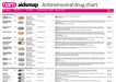 Antiretroviral drug chartA one-page reference guide to the anti-HIV drugs licensed for use in the UK or European Union, with information on formulation, dosing, key side effects and food requirements.
 Your next stepsA booklet with information for people who’ve just found
out they have HIV.
Your next stepsA booklet with information for people who’ve just found
out they have HIV.
 Coming soon: news from IAS 2019 05 July 2019The 10th International AIDS Society
conference (IAS 2019) is being held...
Coming soon: news from IAS 2019 05 July 2019The 10th International AIDS Society
conference (IAS 2019) is being held... Those who are not seen are not heard 01 July 2019A commentary on Optimising the Impact of Key Population Programming...
 The fight for access to abortion care in Europe 27 June 2019Access to
sexual and reproductive health and rights includes safe and...
The fight for access to abortion care in Europe 27 June 2019Access to
sexual and reproductive health and rights includes safe and...