Methamphetamine (‘crystal meth’) exacerbates the HIV-mediated damage to brain cells, according to research published by American investigators in the December 15th issue of the Journal of Acquired Immune Deficiency Syndromes.
The researchers from La Jolla, California found that the toxic effects of HIV proteins on brain cells are worsened by crystal meth in patients with HIV encephalitis, the progressive brain damage that affects movement, memory and verbal fluency. This leads to the destruction of a specific subset of ‘interneurones’ in the frontal cortex.
"When combined, HIV and methamphetamine may work in concert to damage particular non-dopaminergic neuronal populations in the cortex," state the researchers, "enhancing brain metabolic disturbances observed in...HIV-positive methamphetamine users."
However, the investigators also saw that HIV encephalitis was less common and less severe in methamphetamine users, but they fail to reconcile this finding with their observation of enhanced damage to brain cells in those who used the drug and had encephalitis.
Despite this, their results suggest a synergistic relationship between HIV proteins and methamphetamine on the loss of a specific cell population. However, understanding of the relationship between interneurone loss and HIV encephalitis will require further experimental work.
Study findings
In their post mortem study, the group of researchers examined the medical histories of 77 AIDS patients, 34 of whom had regularly used methamphetamine for at least three years prior to death. Both groups had similar levels of opportunistic infections and brain tumours, with the main cause of death being pneumonia or septicaemia.
Although they found fewer cases of HIV encephalitis in the methamphetamine group (9 of 34, 26%) than the non-methamphetamine group (24 of 43, 56%), examination of the HIV protein gp41 in the brains of these patients revealed lower levels in the methamphetamine group (P < 0.05), suggesting that their encephalitis was less severe than in the non-drug group.
Paradoxically, the investigators found evidence of greater damage to brain cells in the methamphetamine users. Although this may be related to the fact that more methamphetamine users showed signs of ‘ischaemic’ or stroke-like damage (7 of 34, 21% vs. 1 of 43, 2%), this finding appears to be at odds with the group’s discovery of less severe HIV encephalitis in methamphetamine users.
Brain cell damage was assessed using antibody-based staining techniques to observe the microscopic structure of the frontal cortex, the region of the brain found behind the forehead that is important in reasoning, planning and abstract thought. In common with previous studies, the researchers saw that brains from patients with HIV encephalitis showed a significant loss of neurones compared with those who had not suffered from the condition. This loss was marginally increased in methamphetamine users, with observed reductions in the number of synapses, the specialised points of contact between neurones.
In an attempt to identify the type of neurone damaged by the combination of HIV encephalitis and methamphetamine, the researchers stained the brain tissue with markers of interneurones, the brain cells that pass signals between neurones within a particular brain region. They saw that the number of interneurones in encephalitis patients was reduced in comparison with those without encephalitis, with those interneurones that remained appearing shrunken, distorted and disorganised.
Methamphetamine only caused further damage to one class of interneurone, which expresses the calcium-binding protein ‘calbindin’ and is ‘non-pyramidal’ in shape (P < 0.05). This led the authors to suggest that this subpopulation is specifically damaged and eventually destroyed by methamphetamine in patients with HIV encephalitis.
The group sought to confirm this finding by examining the effects of HIV encephalitis and methamphetamine use on ‘glia’, the cells that surround and support the electrically active neurones in the brain. An increase in the number of glial cells or ‘gliosis’ is a general response of the brain to all types of damage, including the loss of neurones. As expected, the researchers found that the brains of the HIV-positive patients exhibited a greater degree of gliosis than those of 19 HIV-negative controls, as a consequence of the toxic effects of HIV on brain cells.
Glial cells are found in two anatomically distinct populations: star-shaped astroglia and smaller microglia. By staining these populations separately, the research group found that, although astroglia were unaffected by methamphetamine use, both HIV encephalitis and methamphetamine stimulated further increases in the numbers of microglial cells (P < 0.05). This indicates that having encephalitis and taking crystal meth both worsen the brain damage caused by HIV infection itself.
The investigators propose that crystal meth and HIV proteins could engage the same signalling pathways within these calbindin-positive non-pyramidal interneurones. This could upset the balance of calcium levels, leading to cell damage and death, possibly via effects on mitochondrial activity.

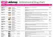 Antiretroviral drug chartA one-page reference guide to the anti-HIV drugs licensed for use in the UK or European Union, with information on formulation, dosing, key side effects and food requirements.
 Your next stepsA booklet with information for people who’ve just found
out they have HIV.
Your next stepsA booklet with information for people who’ve just found
out they have HIV.
 Coming soon: news from IAS 2019 05 July 2019The 10th International AIDS Society
conference (IAS 2019) is being held...
Coming soon: news from IAS 2019 05 July 2019The 10th International AIDS Society
conference (IAS 2019) is being held... Those who are not seen are not heard 01 July 2019A commentary on Optimising the Impact of Key Population Programming...
 The fight for access to abortion care in Europe 27 June 2019Access to
sexual and reproductive health and rights includes safe and...
The fight for access to abortion care in Europe 27 June 2019Access to
sexual and reproductive health and rights includes safe and...