“There’s a wide spectrum of disease that comprises childhood tuberculosis,” said Professor Peter Donald of Stellonbosch University at the Union World Lung Conference in Paris in October.
Most children with TB will present with classical symptoms as chronic cough, fever and weight loss and/or failure to thrive. But, as noted above, after primary infection and the development of the primary complex, the manner in which TB spreads in the body reflects different pathological processes and can have important prognostic significance.7 It can also determine what sort of symptoms the child presents with.
TB can disperse widely through the body in children to many organ systems, and extrapulmonary TB (EPTB) accounts for around 20-30% of TB in children.8 The most serious site of involvement is probably the central nervous system, but TB can also affect the peripheral lymph nodes (especially cervical), intra-abdominal areas, sometimes the ear and the eye, the genitourinary tract (including the kidney), bones and joints, the spine and the skin.9 Although fever and weight loss/failure to thrive are still common in children with extrapulmonary TB, the most conspicuous symptoms (or their primary complaint) may rather be associated with the site of disease.
The following section presents these in order of pulmonary TB, EPTB disease that can be observed on a chest x-ray (intra-thoracic disease), and EPTB elsewhere in the body.
Pulmonary TB
Most children with TB — including those with HIV —have pulmonary TB of one form or another.10 Even within the lungs, however, there can be a number of pathological processes at work.11 These include the early primary infection-related conditions described above, such as ‘uncomplicated Ghon focus,” the localised pneumonic initial infection site and the primary complex, or the Ghon focus with uncomplicated disease in the associated lymph nodes.
“Uncomplicated lymph node disease, in particular hilar adenopathy is the most common finding and is usually regarded as the hallmark of primary tuberculosis,” Marais et al wrote in 2006.12 Although conventionally treated as active disease, in some cases, hilar adenopathy is transient.
Furthermore, Marais et al noted that the conditions associated with primary TB infection, including hilar adenopathy, can show up on x-ray even when there are no symptoms of disease— and many children with these signs will not progress to active disease/primary TB.13 However, given how rapidly TB can progress in children with HIV, even early signs or asymptomatic TB merit aggressive action.
Much early disease, though, according to Professor Donald can be “adequately treated with a touch of isoniazid” (monotherapy, see below) — “though this would certainly not be appropriate when you get into extensive disease manifestations with cavitation and so on.”
“Progression to disease is indicated by the onset of persistent, non-remitting symptoms,” wrote Marais et al. Such disease includes the following: (Citations for the following pulmonary disease section come from several papers from Marais et al)14, 15, 16
- Complicated Ghon focus: in young or immune-compromised children, the MTB infection within the Ghon focus may be poorly contained, leading to necrosis and caseation (the dead tissue decays into a cheese-like mass) which can break through, and cause a cavity to form in the bronchus, potentially leading to endobronchial spread of the infection. Cavitation can also occur, especially in HIV-infected children.17
- Complicated lymph node or lymphobronchial disease: in small children especially, pathology in the lymph nodes can lead to serious complications and bronchial involvement. For instance, swollen lymph nodes, or oedema surrounding the bronchial passage, or polyps within the airway lumen, may partially or completely obstruct the airway, potentially leading to hyperinflation and/or collapse of the lung. Or the diseased lymph nodes may ulcerate and discharge material that can become aspirated, triggering an acute hypersensitivity reaction (epituberculosis), or spreading infection to surrounding airways (infiltrating the parenchyma (lung tissue) — patchy consolidation) or to distal parenchyma in the lungs (expansive consolidation).
- Adult type disease is seen more in adolescence, reflecting a more exuberant immune response that causes significant damage of the lung with progressive cavitation. Ironically, bacilli flourish within these cavities, and TB spreads endobronchially. In contrast to primary TB, adult type disease is often smear-positive for acid- or alcohol-fast bacilli (AFBs, see below).
Pleural TB
The pleura, the membrane that enfolds the lungs, can become diseased either due to the direct spread of caseous material from the lungs or lymph nodes — which can trigger an inflammatory response causing the formation of generally unilateral pleural effusions (pockets of sometimes bloody fluid containing few bacilli). Pleural effusions are less common in younger children. Bilateral or extensive pleural effusions signal more severe disease.
Less commonly, the pleura can become infected through the blood stream —leading to active replication of bacilli and caseation, and the formation of thick bacilli-containing pus within the pleural space (empyema).18
Either form of pleurisy can cause severe chest pain and make it difficult for the child to breathe, but a child with empyema may be more acutely ill.
Pericardial TB
TB can cause inflammation of the fibrous membrane surrounding the heart and effusions after blood borne dissemination or after infected subcarinal nodes ulcerate and discharge caseous material into the pericardial space19. Pericarditis is often life-threatening with cardiovascular symptoms including chest pain, difficulty breathing/shortness of breath, and swollen ankles or legs, dizziness and weakness, enlarged heart, rapid heart beat, heart murmur or sounds, distended jugular veins and pressure, etc.20
Miliary TB
When MTB bacilli spread via the blood stream, they can become lodged within small capillaries where they form tubercles. These may be dispersed throughout the body — affecting different organs and bones — but they more likely to be observed on chest x-rays as diffuse ‘millet-seed”-sized spots.21 In children with HIV, this pattern may be hard to distinguish from lymphocytic interstitial pneumonitis (LIP) (however LIP is associated with generalized symmetrical lymphadenopathy rather than unilateral lymphadenopathy, swollen parotid enlargement and finger clubbing).22 In addition, miliary TB is more common under 2 years of age, and LIP in older children.
Miliary TB is strongly associated with constitutional features (fever and weight loss) rather than respiratory symptoms — but symptoms also reflect the organ systems that are affected.
Central nervous system (CNS) TB
Dissemination of TB to the central nervous system leads to the most dangerous complications of all. Children may develop tubercles and tuberculomas (larger nodular lesions) in the CNS. If one of these ruptures in the subarachnoid space, it can trigger severe inflammation and TB meningitis.23 About half of the children who develop TB meningitis die or have long-term neurological consequences (the management of TB meningitis was covered in more detail in HATIP #98).24
Peripheral tuberculous lymphadenopathy
Swollen peripheral lymph nodes, especially the cervical lymph nodes, are quite common in childhood TB. As opposed to the generalised lymphadenopathy that is often found in HIV, tuberculous lymphadenopathy is usually localised and unilateral.25 TB lymphadenopathy can start out with firm discrete nodes that become matted as the condition develops; eventually there may be granuloma formation, with subsequent caseation or purulent inflammation at which stage the nodes become fluctuant.26 Finally, abscesses and sinuses form leading to drainage, followed by healing and scarring. Until ulcers or fistula form, TB lymphadenopathy is non-painful.
Abdominal TB
TB can also involve the mesenteric or retroperitoneal lymph nodes and the peritoneum, the membrane lining the abdomen, leading to abdominal distension and TB ascites (peritoneal effusions).27 The swollen lymph nodes may adhere to the bowel, causing blockage or fistula formation.
Abdominal TB can cause symptoms such as fever, marked wasting and also abdominal pain.
TB can also involve the colon, possibly as a result of swallowing infected sputum, obstructing the bowel and causing fistula. Again, this is associated with fever and weight loss but there may also be chronic diarrhoea and abdominal pain.
Genitourinary TB
TB can also cause serious renal disease — potentially destroying each kidney if left untreated. Symptoms, which may not be recognised until too late, include difficulty or pain on urinating, blood in the urine, or flank pain. It can also move into the male and female genital tract.
Osteo-articular TB
Bone and joint TB occurs in 1% to 6% of all cases of tuberculosis. Infection of the spine, hips and knees are most common, but infection of other joints may occur. Chronic swelling and pain of the affected joint, without systemic symptoms is common. Tuberculosis commonly affects a single joint, whereas arthritis seen in HIV-positive children more often presents as polyarthritic juvenile idiopathic arthritis or as a spondyloarthopathy (arthritis affecting the spine).28
Spinal involvement (spondylitis/Pott disease), begins in an intervertebral disc and spreads along the anterior and longitudinal ligaments before involving the adjacent vertebral bodies. Abscesses and spinal cord compression and gibbus (spinal deformity humps) may occur – any child presenting with a gibbus should be suspected of having extrapulmonary TB.29 As with other forms of TB, this will be associated with fever, night sweats and weight loss, but also causes stiffness, back pain and difficulty walking, with weakness in the legs and if left untreated, paralysis.
Chronic otorrhoea and mastoiditis
TB has also been reported to infect the middle ear, causing chronic otorrhoea.30 From here, it can spread to the mastoid bone of the skull, potentially leading to the deterioration of the bone structure. Otorrhoea and mastoiditis are associated with ear pain, fever, headaches, and irritability.
TB-related skin lesions
Disseminated TB can also cause skin lesions, such as lupus vulgaris (painful nodular skin lesions usually on the face around nose, eye lids, lips, cheeks and ears) or papulonecrotic tuberculids.
MDR-TB
With the increase in multi-drug resistant (MDR) and extensively drug resistant TB (XDR-TB), there will likely be an increase in drug resistant TB in children. However, Schaaf et al reported no difference in the initial clinical features in children with culture-confirmed drug-susceptible and drug-resistant tuberculosis.31 What clearly sets drug resistant TB apart is its failure to respond to treatment, which can have devastating consequences for children, especially those with the most serious forms of TB such as TB meningitis.32
Congenital TB
Although rather rare — there are only a few hundred cases mentioned in the medical literature — MTB can sometimes be transmitted through the blood or transplacentally via the haematogenous route, or by in utero aspiration of infected amniotic fluid, or during labour from a mother to her infant — and there are numerous reports that this may be more common in the context of HIV.33, 34 The clinical presentation of TB in these infants is somewhat distinct, and merits mention.
Firstly, the site of the primary lesion is not necessarily in the lungs — it may be in the liver or gastrointestinal tract. (The emphasis of the disease may be abdominal or disseminated disease if the infection was spread transplacentally, but if the child breathed in infected amniotic fluid in utero, or infected material during the birth process, it may first lead to pulmonary disease). Generally, symptoms appear within the first few weeks of life, and most commonly include fever, low weight gain, irritability, respiratory distress, poor feeding and marked hepatosplenomegaly.35
Dr Gary Reubenson of Coronation Hospital in Johannesburg described a case of congenital TB during a panel discussion held earlier this year at the South African TB Meeting by the South African Clinician’s Society.
“A newborn baby boy was admitted to the hospital at birth, initially with mild respiratory distress that settled gradually over the course of the next few hours. The mother was HIV-positive and not on ART at that time. But the child had had no postnatal contact with its mother because the very same day that the child was born, the mother was admitted to a TB hospital,” he said.
Over the next two weeks in the ward, the child’s illness progressed, and he developed hepatosplenomegaly. The hospital took blood, urine, CSF and induced-sputum samples and sent them off for TB culture. One of the cultures would eventually come back positive — though the child was put on TB treatment well before that.
“We have a fair number of congenital TB cases in KZN as well,” said Dr Mo Archary of King Edward VIII Hospital during the discussion.

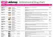 Antiretroviral drug chartA one-page reference guide to the anti-HIV drugs licensed for use in the UK or European Union, with information on formulation, dosing, key side effects and food requirements.
 Your next stepsA booklet with information for people who’ve just found
out they have HIV.
Your next stepsA booklet with information for people who’ve just found
out they have HIV.
 Coming soon: news from IAS 2019 05 July 2019The 10th International AIDS Society
conference (IAS 2019) is being held...
Coming soon: news from IAS 2019 05 July 2019The 10th International AIDS Society
conference (IAS 2019) is being held... Those who are not seen are not heard 01 July 2019A commentary on Optimising the Impact of Key Population Programming...
 The fight for access to abortion care in Europe 27 June 2019Access to
sexual and reproductive health and rights includes safe and...
The fight for access to abortion care in Europe 27 June 2019Access to
sexual and reproductive health and rights includes safe and...